Pablo Irarrázaval Mena, PhD.
Jueves 11 de junio,
09H00 GMT -5
About the Webinar:
Diffusion is the process whereby molecules move in space because of concentration gradients. This displacement involves Brownian motion. If the molecules do not find any obstacles in their movement, the probability of finding them in a given position is determined by a Gaussian distribution. If they do encounter obstacles, as is the case when fibers are present (neurons in the brain, for instance) the probability distribution is no longer isotropic nor Gaussian, and presents preferred directions correlated with the fiber orientations. Knowing this orientation, or even only the diffusivity constants, is the great clinical utility, because they are relevant markers of physiologic activity and functional anatomy.
On the other hand, magnetic resonance imaging (MRI) is sensitive to the spins’ motion (associated to the Hydrogen atom). Using this sensitivity it is possible to design MRI acquisition techniques to measure diffusion. In this talk I will visit the different techniques employed to acquire the diffusion signal. I will start with the simplest technique and work our way to the most complex ones: fundamentals of diffusion acquisition; Diffusion Weighted Imaging (DWI); Anisotropy; Diffusion Tensor Imaging (DTI); and Diffusion Spectrum Imaging (DSI). For this last technique I will introduce the concept of q-space. Finally, I will present some of the applications of the diffusion images, in particular regarding the study of brain connectivity.
About the presenter:
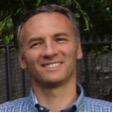
I am fa scinated with the application of signal processing to medical imaging, in particular to Magnetic Resonance Imaging (MRI). I have dedicated most of my working time to research and teaching. I did my undergraduate studies at Pontificia Universidad Católica de Chile, where I received my Electrical Engineering degree (1988) and later on continued at Stanford University where I obtained my M.Sc. (1990) and Ph.D. (1995) degrees. Since then, I have done an academic career in the Department of Electrical Engineering at the School of Engineering of the Universidad Católica, including some administrative positions—Department Chair, Vice-Dean for Undergraduate Academic Affairs, and faculty’s representative in the University Board.
 scinated with the application of signal processing to medical imaging, in particular to Magnetic Resonance Imaging (MRI). I have dedicated most of my working time to research and teaching. I did my undergraduate studies at Pontificia Universidad Católica de Chile, where I received my Electrical Engineering degree (1988) and later on continued at Stanford University where I obtained my M.Sc. (1990) and Ph.D. (1995) degrees. Since then, I have done an academic career in the Department of Electrical Engineering at the School of Engineering of the Universidad Católica, including some administrative positions—Department Chair, Vice-Dean for Undergraduate Academic Affairs, and faculty’s representative in the University Board.
My research career has revolved around Magnetic Resonance Imaging. During my Ph.D. and continuing as a faculty in Chile in the 1990s I worked on k-space trajectories. K-space is how the MRI community refers to the Fourier domain, where the scanner samples the data. My early work was related to devising new ways to traverse the three dimensional k-space for regular imaging. Later on, in the 2000s, I became interested in under-sampled acquisition and its reconstruction techniques. I have worked combining reconstruction with image registration, object modeling, and lately in compressed sensing.
I have been fortunate enough to share with many bright people, with whom we have achieved several goals. I have had more than thirty graduate students, and several research associates and postdocs. In 1999, together with the School of Medicine we founded the Biomedical Imaging Center. Our research has been published in several scientific journals and conference presentations. We have also produced international patents. And finally I am proud that we were able to start up a company that is helping to translate these results to the “real world.”

